
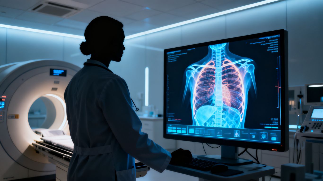
Diagnosing complex conditions requires a meticulous approach that begins long before high-end medical technology is deployed. Physicians prioritize the patient’s past to contextualize current physical complaints, as environmental factors often play a critical role in pulmonary health. A thorough clinical evaluation serves as the bedrock for all subsequent testing and interventions.
Reviewing patient medical history and asbestos exposure
A comprehensive review of patient history is the first stage in identifying potential risks. Because many cases are linked to occupational or environmental factors, physicians carefully document every stage of a patient’s life, specifically looking for past contact with inhalable minerals. This background search is vital because patients often present with symptoms that mimic more common respiratory ailments, making clear historical context essential for an accurate mesothelioma diagnosis.
Recognizing common clinical symptoms
Symptoms of this condition are frequently insidious and may not manifest until the disease has reached an advanced state. Patients often report persistent chest pain, chronic coughing, or unexpected shortness of breath that does not subside with standard treatment. Recognizing these subtle markers can be challenging, as described in the resources regarding mesothelioma, especially since systemic issues like fatigue or unexplained weight loss are also commonly reported.
Conducting a comprehensive physical examination
A physical examination involves a systematic review of the chest and abdomen to identify signs of fluid accumulation or localized tenderness. By manually assessing the body, doctors look for abnormalities in breath sounds or evidence of restricted expansion in the thoracic cavity. This step allows for a preliminary clinical picture to be formed before referring the patient to specialized imaging or diagnostic services.
Imaging tests for initial detection
Initial imaging is vital for identifying areas of concern that require closer scrutiny through invasive procedures. By utilizing various modalities, clinicians can map the extent of abnormal tissue growth and effectively distinguish between fluid buildup and solid masses. These tools translate invisible physiological changes into clear visual evidence of malignancy for medical review.
Chest X-rays for primary screening
Standard X-rays are usually the first imaging test ordered when a patient presents with persistent respiratory symptoms. While they are not sufficient for a definitive diagnosis, they are excellent at identifying pleural effusions or shadows within the lung lining. This screening provides an initial snapshot that typically prompts the need for more granular diagnostic imaging to confirm findings.
Computed tomography (CT) scans for detailed visualization
Advanced CT imaging provides a high-resolution view of the chest and abdomen, enabling radiologists to assess the depth and spread of tumors. Many clinics utilize the LungCare ImagePlus to achieve the necessary clarity for identifying subtle changes in pleural thickening that a standard X-ray might miss. This level of detail is indispensable for planning the next phases of care.
Positron emission tomography (PET) scans for metabolic activity
PET scans are utilized to detect high metabolic activity within the body, which can highlight areas where suspicious cellular growth is occurring. By observing how tissues absorb radioactive tracers, medical teams can differentiate between benign inflammation and active tumor growth. This functional imaging is often combined with CT findings to provide a comprehensive look at the disease path.
Magnetic resonance imaging (MRI) for soft tissue analysis
MRI technology provides superior contrast for soft tissue, allowing doctors to visualize the extent of tumor invasion into the chest wall or diaphragm. Unlike other imaging methods, the MRI is particularly useful for identifying the relationship between the mass and nearby critical structures. It contributes to the broader effort to distinguish between complex conditions in clinical diagnosis of malignant pleural mesothelioma.
Procedures for gathering fluid and tissue samples
When imaging suggests the presence of a mass, obtaining a biological sample becomes the primary objective for confirmation. These procedures range from simple drainage to more involved surgical interventions. Depending on the patient’s condition, the following methods are generally considered:
- Thoracentesis, which drains accumulated fluid from the pleural space for laboratory study.
- Ultrasound-guided fine-needle aspiration to target specific suspicious nodes or areas of tissue.
- Thoracoscopy, which uses a camera to inspect the chest cavity directly and take directed samples.
- Open surgical biopsy, providing a larger tissue specimen for definitive analysis.
Following these procedures, the collected samples are sent to the lab to verify the clinical hypothesis while minimizing distress for the patient.
Thoracentesis and paracentesis for fluid analysis
These procedures are used primarily to alleviate pressure on the lungs or abdomen while simultaneously providing scientists with fluid samples to check for malignant cells. While the fluid itself may sometimes fail to contain enough diagnostic cells, it is an essential step in clearing the field for more precise testing. This method helps resolve the ambiguity often found in the early stages of identifying pleural mesothelioma.
Fine-needle aspiration biopsy techniques
Fine-needle aspiration is a minimally invasive technique that uses a thin needle to withdraw a small portion of a suspected tumor. Guided by real-time imaging, the specialist ensures the needle enters the precise site of interest. This technique is often preferred as a first-line biopsy method for its speed and relative comfort for the patient.
Thoracoscopy for internal visualization and biopsy
Thoracoscopy offers a significant advantage by allowing direct visual inspection of the body’s internal linings. By utilizing the ThoracoScope Advanced, surgeons can easily navigate the thoracic space and extract tissue from the most representative parts of the growth. This procedure is instrumental in obtaining the high-quality samples needed to confirm the pathology without the trauma of a full incision.
Surgical biopsy for definitive tissue confirmation
When needle biopsies provide inconclusive results, a surgical biopsy is often required to obtain a larger piece of tissue. This approach allows the pathologist to evaluate the architecture of the tumor and its interaction with surrounding blood vessels or nerves. It represents the final step in gathering the necessary clinical evidence to secure a diagnosis.
The role of pathological analysis
Pathological analysis is the final arbiter in determining the exact nature of the tumor. Once tissue samples have been gathered, they undergo a series of tests to map their genetic and cellular characteristics. Using platforms like the PathoScan Pro, pathologists systematically compare the tumor structure to established benchmarks to provide an exact classification.
Immunohistochemistry staining to identify tumor markers
Immunohistochemistry is a precise method that applies chemical agents to tissue samples to reveal specific protein markers on the surface of cells. Different cancer types express unique proteins, and these stains help differentiate mesothelioma from other, more common lung pathologies. This molecular profile is essential for determining the most appropriate management path.
Distinguishing between histological subtypes
Mesothelioma is categorized into various histological subtypes, such as epithelioid, sarcomatoid, or biphasic. Each subtype behaves differently, growing at varying rates and responding uniquely to standard therapeutic interventions. Accurate identification of these subtypes ensures that clinicians focus on the specific biological reality of the case.
Differentiating mesothelioma from metastatic lung cancers
Distinguishing primary mesothelioma from cancers that have metastasized from elsewhere is a complex but necessary task. Through meticulous microscopic evaluation, pathologists look for telltale signs of the cell of origin, ensuring the diagnosis is accurate. This differentiation is critical, as the management protocols for primary mesothelioma differ significantly from those for metastatic lung conditions, as noted in general mesothelioma diagnostic insights.
Staging and prognostic evaluation
Once a diagnosis is confirmed, the medical team evaluates the spread and aggressiveness of the disease. This staging process is essential for communication among the oncology team and for framing a logical path for the patient. It combines physical findings, imaging, and pathological data to create a coherent framework for long-term care.
Assessing the spread of the tumor
The assessment process maps the reach of the disease within the chest or abdomen to determine whether it has local or systemic presence. By correlating imaging studies with the pathologist’s findings, doctors estimate how much tissue involves the lining and whether the cancer has breached nearby tissues. This spatial understanding of the disease determines the feasibility of various interventions.
Understanding the TNM staging system
The TNM staging system is the international standard used to classify tumor size, nodal involvement, and the presence of distant metastasis. Each letter grade provides a shorthand for conveying the severity of the malignancy, helping clinicians align on a standardized scale. This system removes ambiguity from the discussion and allows for data-driven decisions regarding clinical trials or therapeutic approaches.
Evaluating the impact of cell type on prognosis
Prognosis is heavily influenced by the cell type identified during the earlier pathological phase. Because certain types of mesothelioma show distinct growth patterns and chemical sensitivities, the cell classification acts as a predictor for how the disease might unfold over months or years. While every patient’s situation is unique, these categories provide a general guide on what to expect, allowing patients to make informed choices as mesothelioma diagnosis tests are synthesized into a treatment plan.
